Why Does My Eye Doctor… Part 4

Welcome back to my “Why Does My Eye Doctor…” series!

Have you ever wondered, “Why does my optometrist care so much about my medical history? That has nothing to do with my eyes!!”

If so, you’re not alone.  It can definitely seem weird being asked (or being the doctor and asking) about general medical history while at the eye doctor, but I promise it isn’t just to be nose-y!

Believe it or not, your ocular health is closely related to your systemic health, and your eyes are often one of the first places to show systemic disease!

Sound crazy?  Let’s take a look at four (relatively) common conditions that can be seen in your eyes!

Diabetes

  • Diabetes is a disease that affects the small blood vessels in your body.  (Okay, it affects larger vessels too, but for the sake of the eye, we’re focusing on the smaller ones.)
  • In the eyes (specifically the retina), this can cause bleeding, or hemorrhages, as well as leakage of other materials from the blood vessels.  If this bleeding or leakage happens to be around the macula, or part of the eye responsible for your best vision, it can cause swelling that then distorts your vision.
  • Fluctuations in blood sugar levels can also affect your vision, as they cause changes within the lens of your eye.  If you notice frequent changes in vision, it may be a sign of diabetes.

Hypertension

  • High blood pressure is, once again, a disease of the small blood vessels in your body.
  • The retinal disease (retinopathy) associated with hypertension occurs in 3 main stages.
    • Vasoconstrictive: This is a fancy way of saying that the blood vessels constrict in response to the increase in blood pressure.  The goal of the constriction (which is automatically controlled) is to decrease the amount of blood flow so that the normal vessel system of the retina isn’t overwhelmed.  This early stage is seen in the eye as arteriolar narrowing.  *I would include a picture, but it can be pretty hard to appreciate*
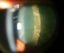
    • Sclerotic: The term sclerosis simply means hardening.  The constriction of the blood vessels in the vasoconstrictive phase occurs via a muscular layer in the arteries.  Like any muscle that is frequently used, over time, this muscular layer gets bigger, making the arteries harder.  As the arteries harden and become heavier, they compress the underlying veins.  This causes a characteristic appearance of blood vessel “nicking” as seen below, where the vein becomes diminished upon artery crossing.
Arteriovenous nicking - Wikipedia
    • Exudative: Once enough compression has occurred in the sclerotic phase, blood, fats, and additional components begin to leak out of the vessels, signaling a transition to the exudative phase.  In this stage, hemorrhages (areas of bleeding), exudates (hard, fatty deposits), and edema (swelling from leaking fluid) are common.  As blood vessel compression also reduces blood flow (stasis), clots are more likely to form, occluding, or blocking the vessel.  Without proper blood flow, ischemia (literally lack of blood flow) occurs, causing subsequent damage to affected tissues. 
This is Grade 3 hypertensive retinopathy. Diastolic pressure is usually  110-115 mmHg and retinal arteries lose their abilit… | Retinal photography,  Eyes, Eye health

Thyroid Disease

  • First things first: thyroid problems can come in two forms – overactive (hyperthyroid) or underactive (hypothyroid).
  • Both versions can be caused by autoimmune states.  The one most commonly associated with hyperthyroidism is Grave’s Disease, while the one most commonly associated with hypothyroidism is Hashimoto’s Disease.
  • In addition to systemic manifestations, both hyper and hypothyroidism may present in the eyes. Possible symptoms include:
    • Redness
    • Dryness and irritation
    • Swelling around the eyes
    • Protrusion of the eyes
    • Pain with eye movement
    • Inability to fully close the eyes
    • Double vision
    • Loss of vision
  • These symptoms may occur in existing cases of autoimmune related thyroid disease, or may be among the first presentations of the condition.  If your doctor suspects thyroid disease secondary to ocular presentations, they will likely ask a number of systemic questions that seem totally unrelated such as:
    • Unintentional/unexplained weight gain or weight loss
    • Sensitivity to heat or cold
    • Changes in energy levels (either very high or very low)
    • Changes in mood (such as being highly irritable or more depressed)
    • Changes in bowel movements
    • Changes in menstruation
  • If your doctor asks these seemingly off the wall questions, don’t freak out!  They’re trying to narrow down the potential causes of your symptoms so that they can send you to the right place to get the right diagnosis!

Multiple Sclerosis

  • While we’re on the topic of autoimmune diseases, it’s important to discuss Multiple Sclerosis.
  • In this condition, antibodies attack the myelin (protective coating) that surrounds nerves in your central nervous system (brain and spine).  This causes demyelination (literally, loss of myelin) around the nerve, which impedes transfer of the signal from the brain to its intended recipient.
  • One of the more common, and often early, presentations of multiple sclerosis is optic neuritis, or inflammation of the optic nerve.
    • Optic neuritis generally presents as mild-moderate vision loss in a single eye that comes on suddenly and may worsen for around 2 weeks, after which symptoms begin to improve.  Colors may seem less vibrant out of this eye, and there is often pain with eye movement.
    • As with any sudden decrease/loss of vision, it is important to see an eye doctor with these symptoms.
    • Looking in the back of your eye, the doctor will often see nothing out of the ordinary.  This is because the inflammation is behind your eye (retrobulbar).
    • Your doctor should order an MRI, with and without contrast, to aid in diagnosis.  The results of this test provide important information regarding future prognosis as:
      • 1/4 people with no other lesions on MRI have a diagnosis of Multiple Sclerosis within 15 years
      • 2/4 people with optic neuritis are diagnosed with Multiple Sclerosis within 15 years
      • 3/4 people with another characteristic MRI lesion are diagnosed with Multiple Sclerosis in 15 years
  • As before, your eye doctor may ask about other systemic symptoms as well, such as:
    • Fatigue
    • Dizziness
    • Numbness/Weakness
    • Tremor
  • Please don’t avoid the questions, or think your eye doctor is practicing outside his or her scope!  Believe it or not, as optometrists, we are trained to know the symptoms of systemic diseases that may present in your eye.


If you learned something from this post, please share it with a friend or family member!  If you liked it, please subscribe, or like my page on Facebook! And as always, if you have any questions or comments, please contact me – I’d love to hear from you!

This is Part 4 of my “Why Does My Eye Doctor” series – click the links below to check out Parts 1-3!

Part 1 – Dilation
Part 2 – Which is Better, 1 or 2?
Part 3 – Air Puff Test

Optometry in Focus: Cataracts

Happy Wednesday, y’all!

Since it’s the middle of the week, how about we start out easy:

How many of you know someone (either a friend or a relative) who has had cataracts?

Hopefully, unless you live under a rock or are under the age of 15, you all know of at least one person who has, or has had, cataracts.

Surprisingly, even though most of the world knows of someone with cataracts, few people seem to know of what they are, or what they mean for you and your vision.

Case in point: one of the optometry groups that I follow on social media posted yesterday how they had just finished telling a patient that he had cataracts, only for him to report that they told him he had cancer!

While this may seem like an extreme interpretation, I’ve had many patients who are convinced cataracts are equivalent with a visual death sentence (and also the occasional few who are convinced they have a “Cadillac” in their eye…)

Considering the number of misconceptions that exist, I’ve decided to use today’s post to discuss cataracts, and hopefully dispel some common myths!

Let’s get started.

Cataracts in Focus

First things first (someone should start tallying up how often I say that…): ocular anatomy.

Eye Health: Anatomy of the Eye - VisionAware 

Okay, in the lovely picture above, you can see all the major components of the eye.  Our focus for today is the lens, which sits right behind the iris, or colored part of the eye.

The role of the lens in your eye – like every lens really – is to focus light to create a clear image.

However, unlike lenses for your camera, or lenses in your glasses, or contact lenses that you put on your eyes, the natural lens of your eyes is made up of cells, proteins, and fibers!  Together, these components come together to form distinct layers of the lens (as shown below).

Because of its cellular makeup, your natural lens changes over time – adding layer after layer through the years!

Unlike other parts of your body, however, the natural lens of your eye is sanctioned off from the rest of the eye by the capsule – which is essentially an elastic bag that holds the lens (epithelial) cells and lens fibers in place.  While this layer does an excellent job at protecting the lens from viruses and bacteria, it also prevents any of the layers from being lost.  In other words, the lens fibers that you’re born with are still in your eye when you’re 50!

Which is all well and good, until something causes a structural change in either the lens cells or lens fibers, resulting in opacification (clouding) of the lens.

Aka: a cataract

Cataract Causes

Cataracts have approximately 8,000 different causes.

Okay, this is an exaggeration, but there are many different reasons for the formation of cataracts!

Some common ones are:

  • Age
  • Trauma
    • Direct trauma to the lens
    • UV Radiation
    • Ionizing Radiation (think cancer treatment)
  • Diabetes
  • Systemic steroid use
  • Poor nutrition
  • Alcohol use
  • Smoking

Interestingly, different causes of cataracts result in different types of cataracts!

Nuclear Cataract

Age-related cataracts. Immature cataracts (A, B, C) and mature cataract...  | Download Scientific Diagram

Nuclear cataracts are one of the most common type of cataracts.  This type of cataract originates in the central part (nucleus) of the lens, and is most commonly associated with increased age. What initially starts out as a browning (brunescence) with slight clouding, can eventually become the milky white cataract that you see in the bottom right picture!

Cortical Cataract

Cortical | Columbia Ophthalmology

Cortical cataracts are admittedly some of my favorite to see in patients – they can look so pretty!

These cataracts start from the outside and gradually work their way in in a spoke-like pattern, as shown above.  Cortical cataracts can also be associated with age, but additionally have a higher incidence in patients with diabetes.

Posterior Subcapsular Cataract


As indicated by the name, posterior subcapsular cataracts involve an area of the lens just ahead of the posterior (back) portion of the lens capsule.  This type of cataract generally begins as a small, cloudy area, but eventually enlarges to cover more of the back portion of the lens.  Interestingly, posterior subcapsular cataracts generally have the most visually significant changes over a short period of time – potentially altering vision within just a few months!  Like cortical cataracts, these are more common in patients with diabetes, however, they are also associated with steroid use, a highly near-sighted prescription, and some ocular diseases.

Cataract Symptoms

Despite there being many different causes and types (I only listed the top 3) of cataracts, most people report similar symptoms:

  • Glare/halos (especially when driving at night)
  • Decreased vision (that is not improvable with glasses)
  • Potential yellowing of vision

Cataract Treatment

Chances are (if you’re still reading at this point) that you’re thinking some variant of:

“That’s great, Hannah.  But, what I really care about is what to do about them!”

Unfortunately, as it currently stands, there is only one form of treatment for developed cataracts: surgery.

Which is normally the part where people freak out, and I get it!  I’m personally not particularly thrilled by the notion of any type of surgery.  However, before your amygdala grabs you, let’s take a moment to discuss cataract surgery.

First up: cataract surgery is the most commonly performed ocular surgery, and one of the most common surgeries in the world! (I tried to find specific stats, but came up somewhat empty handed.  In 2015, over 20 million cataract procedures were performed worldwide, with somewhere around 3.6 million in the US alone.)

Being performed so frequently, the risk of complications is also impressively low.  Approximately one percent of patients may experience minor complications or side effects (ie dry eye), while only around 1/1000 may experience more major complications that causes longstanding reduced vision.

In this outpatient procedure, your cataract surgeon removes the natural lens from your eye (while leaving part of the capsule intact if possible) and inserts a new, synthetic lens that is calculated to fit your visual needs in its place!

Sometime after the initial cataract surgery, many people end up getting a “secondary cataract”, when some remaining lens cells migrate and cover the intact capsule.  Thankfully, this condition is also easily treated; your ophthalmologist (or optometrist in a select few states) simply uses a laser to open up the capsule and restore clear vision!


There are many more topics related to cataracts and cataract surgery that could be discussed, but for today, I’ll call it good!   If you have any questions or comments, please contact me – I’d love to hear from you!

Likewise, if you learned something from this post, please share it with a friend or family member!  If you liked it, please subscribe, or like my page on Facebook!

Pediatric Eye Exams: How Does My Doctor… Part 3

Welcome back for Part 3 of my Pediatric Eye Exam series!

*Click here for Part 1 and here for Part 2*

In the first two posts of this series, I discussed how to assess visual acuity in an infant and how to do some basic additional testing.

Today, however, I’m gonna jump into one of the most common questions that I get:

How do you determine the glasses prescription of an infant?! They can’t tell you which is better one or two!


Refraction, in an infant or young child, is admittedly a much different procedure than in an adult.

As I discussed in my second post of the “Why Does My Eye Doctor” series, refraction in an adult typically involves showing a variety of lenses in a phoropter, and asking which one is subjectively better.

In infants and young children, however, refraction is done in free space, using retinoscopy.

What does this mean?

First things first, retinoscopy is a procedure by which the eye doctor shines a light towards the back of the eye and observes the ensuing reflex.  Lenses then may be added to try to neutralize, or minimize, the movement seen.  The lens that corresponds to the least amount of reflex movement is recorded as the prescription.

Free space just means that this procedure can literally take place anywhere!  Since most infants and young children are not particularly skilled at sitting still, this is a vital component.  With loose lenses, it is possible to much more easily follow the child as he or she moves around the chair/room.

However, as infants and young children can’t necessarily be trusted to focus on a specific target, dilating drops are often used to paralyze the focusing system to ensure that there isn’t fluctuation in the prescription found.

As discussed in my post, Decoding the Numbers: Hyperopia, most infants are far-sighted when they are young, with some amount of astigmatism.  However, just because a prescription is found does not mean that full, or even part-time glasses wear is necessary.  Rather, lenses are only prescribed when the prescription falls outside of the normal bounds, posing a potential for reduced vision, or amblyopia.

When glasses are prescribed, though, since infants and young children often can’t vocalize visual improvements, they are followed more frequently – often at 6-8 week intervals.  At these visits, vision and prescription are reassessed to determine prescription accuracy and visual improvement.  Once the accuracy has been confirmed and visual stability achieved, the child may then return to a normal follow-up pattern.

And there you have it!

If you learned something from this post, please share it with a friend or family member!  If you liked it, please subscribe, or like my page on Facebook! And as always, if you have any questions or comments, please contact me – I’d love to hear from you!

Contact Lenses: What Are My Options? Part 2

Hello all, and welcome to Part 2 of my Contact Lenses: What are My Options? series!

 *Click here for Part 1!*

In the last post, I began the discussion on soft contact lenses, and provided some (okay, probably too much) information regarding lens material and the benefits of each type.

Today, I’m going to stick with the soft contact lens theme, but switch away from the material conversation, and focus instead on lens replacement schedule.

Let’s get started – I promise it’s not as boring as it sounds!


Soft Contact Lenses: Lens Replacement

Quarterly

Basics:

  • This lens is to be worn daily for 3 months.
  • Quarterly replacement lenses are relatively uncommon, and are most commonly specialty lenses, reserved for high prescriptions, or other ocular conditions.
  • Like all lenses, it should be removed for sleeping, and stored in a contact lens case in contact lens solution.
    • Lens case should be replaced every 3 months.
    • Contact lens solution should be dumped daily, and filled with new solution.
    • For optimum comfort, health, and vision, lenses should be manually cleaned (aka scrubbed with the tip of your finger) to help remove debris and deposits.
  • If you are in a quarterly lens, chances are it’s because that’s one of the only soft lens options for you, which is a pro, and a con.

Monthly

Basics:

  • This lens is to be worn daily for a month.
  • Like all lenses, it should be removed for sleeping, and stored in a contact lens case in contact lens solution.
    • Same details as above.

Pros:

  • Normally, monthly lenses are some of the more cost effective lenses.
  • Less waste (packaging, lenses, etc)

Cons:

  • Prone to overuse (wearing them longer than a month)
  • Prone to allergic responses
  • Prone to deposits
  • Increased potential for complications (secondary to overuse and improper cleaning/storage)
  • Added expense of contact lens case and solution

2-Week

Basics:

  • This lens is to be worn daily for two weeks.
  • Like all lenses, it should be removed for sleeping, and stored in a contact lens case in contact lens solution.
    • Same details as above.
Pros:
  • It’s replaced more than a monthly lens, decreasing some of the potential for allergic responses, deposits, and other complications.
  • Two week lenses may still be cheaper than daily lenses.
  • Less waste than dailies

Cons:

  • Even more prone to overuse (two week lenses often become monthly lenses)
  • Prone to allergic responses
  • Prone to deposits
  • Increased potential for complications
  • Added expense of contact lens case and solution

Daily

Basics:

  • This lens is worn for a single day, then disposed of.
  • Daily lenses are not meant to be slept in.

Pros:

  • Low risk profile
  • Improved comfort and vision
  • Overuse is rare
  • No (okay, minimal) need for contact lens cases or solution

Cons:

  • Potential increased cost upfront (though balanced out by reduced expenditures on solution)
  • Increased waste (lenses and packaging)


Personal Preferences

One of the best parts of optometry school – at least for a moderate myope – was getting to tryout most any lens that I was interested in.  As such, I’ve tried most lens replacement schedules.

Daily lenses are hands down my favorite, as, from my experience, they provide the best comfort and vision out of any soft contact lens replacement schedule.  There’s no concern of having enough solution with me when I travel, and no disgusting looking contact lens cases.  They’re lighter (they only have to last a day!), and leave my eyes feeling like they can breathe!

I additionally love their decreased risk profile.  As a moderately near-sighted individual, I completely understand patient’s frustration in being told to never use contacts around water, be it showering, swimming, or any other water related activity.  With daily lenses, I am more comfortable saying – you can keep wearing your lenses, just make sure to replace them after you finish the activity.

As much as I love them, daily lenses aren’t always the best option for every eye.  Sometimes the prescription is too unique, and requires a longer use lens.  Other times, the specific lens capabilities (ie Transitions contact lenses) aren’t offered in daily lenses.  Whatever the case, rest assured that your optometrist is committed to providing you with the best comfort, health, and vision that they can!

If you learned something from this post, please share it with a friend or family member!  If you liked it, please subscribe, or like my page on Facebook! And as always, if you have any questions or comments, please contact me – I’d love to hear from you!

Contact Lenses: What are my options? Part 1

In this day and age, when most people think of contact lenses, they immediately think of soft contact lenses – and rightfully so! Soft contact lenses are the most frequently prescribed lenses, accounting for over 80% of the contact lens market.
But did you know that there are other types of contact lenses?
Or that all soft contact lenses are not made equal?
If not, keep reading as I answer the question:

Contact Lenses: What Are My Options?

When we’re discussing contacts, there are three major categories: soft contact lenses, hard contact lenses, and hybrid contact lenses. In today’s post, I’m going to dive into the lens everyone knows and loves – the soft contact lens!

Soft Contact Lenses

Soft contact lenses, for a simple definition, are flexible lenses that sit on the eye to provide refractive correction.  These can be divided into several different categories, based on material and wear time.  Let’s start with material.
Soft contact lenses can be hydrogel or silicone hydrogel.
What’s the Difference?
Hydrogel lenses are made from a gel-like plastic substance that is very good at holding water (hence hydro-gel). This is what the very first soft contact lenses were made of, and makes for relatively comfortable lenses that allow some oxygen to pass through.
Silicone hydrogel lenses are similar to hydrogel lenses, however, they have an additional component – silicone.  Adding silicone to the initial hydrogel material allowed for improved oxygen flow through the lens, however, it decreased the ability of the lens to hold water.
Why Does It Matter?
With hydrogel and silicone hydrogel lenses, there are two main factors that we’re looking at – water content (how much water the lens can hold), and oxygen permeability (how much oxygen can pass through the lens).  Both are important for maintaining the health of your eye.
First up, water content.
 
Okay, I’m gonna start this part out a little weird.
 
Stop what you’re doing, and look at your skin.

Is there water on it? (The answer should be no unless you just got out of a body of water or are actively sweating..)

Is it uncomfortable?  Chances are the answer is also no.
Why not?

Because the skin (or the outer layer of it anyway) that covers your arms, legs, etc, is keratinized – which just means that it has a material (keratin) in it that helps to form a hard(er) protective barrier

Now, take a look at your eyes – they obviously look quite a bit different, right?
Though there’s a lot more to it, one of the major differences is that the outer layer of tissue in your eyes doesn’t have keratin!  It’s missing part of that protective barrier!
While this is a great thing for vision (keratinized skin doesn’t exactly promote sight…), it means that your eyes need something else to stay happy (comfortable) and healthy (alive and transparent) – moisture!
 
From that point, the water content (how much moisture it holds) of your contact lens is important, as it helps determine the comfort, clarity, and consistency of your vision!
 
With this information in mind, it would probably be easy to say: give me the contact lens with the highest water content!
 
Unfortunately, water content and oxygen permeability are inversely proportional: the more water is in your lens, the more difficult it is for oxygen to pass through.
 
But why does oxygen matter?
 
Let’s go back to high school or college biology for a bit.

Your body is made up of cells – which are made up of even smaller components called organelles (tiny organs).  Each of these organelles has a role in the functioning of the cell factory.  You’ve got the foreman (nucleus), the assembly line (endoplasmic reticulum), the packaging plant (golgi apparatus), even the janitor/maintenance crew (lysozome).  But how is this all powered?  Oh yeah, by the energy source: the mitochondria.

Keep your thinking caps on, we’re not done yet – now we get to fast forward a few lectures to cellular respiration day.
Cellular respiration is the process by which cells (well, specifically the mitochondria) take simple sugars (glucose) and break them down to create energy (ATP).  This process can be broken down into 4 (not so simple) steps:
  • Glycolysis (Glucose -> pyruvate, ATP, NADH)
  • Pyruvate Oxidation (Pyruvate -> Acetyl CoA, NADH, CO2)
  • The Krebs/Citric Acid Cycle (Acetyle CoA -> ATP, CO2, NADH, FADH2)
  • Oxidative Phosphorylation (NADH + FADH2 -> H2O + ATP)

The catch? The last three of these steps require oxygen.

Which is pretty much to say: for your cells to (efficiently) produce energy, they need oxygen!  And that oxygen is carried by the blood.

Take a look at your eyes again – paying particular attention to your (clear) cornea).  Do you see any blood vessels?

Hopefully the answer is no.

To promote the transparency that allows for vision, the cornea is naturally avascular: it doesn’t have blood vessels!  This means that it has to get its oxygen from somewhere else – the air. However, when you put a contact lens on, that ability to take in oxygen from the air is decreased.

Without proper levels of oxygen, the cornea can swell or, in cases of chronic oxygen depletion, form new, leaky blood vessels that may eventually cause permanently reduced vision.


So then, what’s the solution?

Honestly, it’s all about finding the balance that works best for your eyes, as determined by your prescription, wear time, and pre-existing conditions (like dry eye), and not being afraid to try a new lens if your current ones aren’t working!

In the grand scheme of things, today’s discussion barely scratches the surface of all there is to know about contacts, so be sure to stay tuned for a later post to learn more about your lens options!

If you learned something from this post, please share it with a friend or family member!  If you liked it, please subscribe, or like my page on Facebook! And as always, if you have any questions or comments, please contact meI’d love to hear from you!

Optometry Do’s and Don’ts

Hey guys – it’s Fun Post Friday!

Fun posts are probably my favorite way of rounding out the work week, and what better topic than some simple optometry do’s and don’ts!  Let’s get started!

Do’s

  • Wear your sunglasses! UV-blocking sunglasses help protect your skin and eyes from potentially damaging ultraviolet light, promoting better ocular health with age! 
  • Have a dilated eye exam every 1-2 years! You can read an in-depth discussion of this in my first post of my “Why Does My Eye Doctor…” series.  In summary though, dilated eye exams are vital – not only for monitoring potentially asymptomatic ocular conditions, but also in diagnosing and managing systemic conditions.
  • Ask questions! If you’re ever unsure of something that your eye doctor has told you, or have questions about something that you’re experiencing, let them know!
  • Wash your hands before touching your eyes!  This sounds like a no-brainer, but good hygiene is important for optimal ocular health (even when we’re not in the middle of a global pandemic)!
  • Wear safety glasses! While taking out foreign bodies (ie anything that is stuck in/on your eye that isn’t supposed to be there) can be exciting as an optometrist, it isn’t fun for you! Plus, some foreign bodies can be visually devastating.  It’s always better to be safe than sorry!
  • Eat a healthy diet! I know, I know… why am I, as an optometrist, talking about food?!  Because it matters for your eyes! For the sake of time, I won’t dive into specifics today, just know that the same foods that are important for keeping your body strong are vital in eye health!  If you’re interested in learning more, keep your eyes peeled (pun intended) for an upcoming post in which I talk about diet and eyes!

Don’ts 

  • Sleep in your contacts!*  This is one that I remember being told the first day that I was fit in contacts – like hopefully all of you who wear contacts were.  Why not?  Because sleeping in contacts significantly increases your risk of eye infection.  Sleeping (without contacts) naturally decreases the amount of oxygen and hydration to the cornea (clear surface of the eye).  However, when you add a contact lens to the mix, the oxygen and tear-layer are further decreased, reducing your eye’s ability to fight off potential infections!
    • For those of you who may argue that you’ve slept in contacts before and never had problems – great! I’m glad you’ve been lucky so far.  However, just because it hasn’t happened to you, doesn’t mean it can’t or won’t, and the visual results of such ocular infections can be devastating.  Don’t believe me? Google it.
  • Use Vizine or other “red remover” drops! I get it, no one likes red eyes.  However, redness is often an indicator of an underlying problem! Trying to get rid of the red may make your eye look better, but it doesn’t address what’s really going on.
    • Why is your eye red? That would be a great…25ish part series? For a quick list of some options though, it could be dry eye, allergies, a bacterial infection, a viral infection, a burst blood vessel, or inflammation of the eye of a number of different causes.
    • If you’ve been to the eye doctor and had all infectious/inflammatory causes of red eyes ruled out, but you still have some redness, still don’t reach for Vizine.  This specific product decreases the red by causing the blood vessels to shrink or constrict. However, after the initial constriction, the blood vessels actually expand or dilate, causing “rebound redness”!
    • The good news though is that there is a drop that decreases the redness without rebound effects!  It’s called Lumify** and it literally decreases the redness in under a minute.  However, just like before, this drop should only be used after the cause of your red eye has been determined!
  • Go to the ER for eye problems!*  Even though I’ve only been around the optometric field for something like 6 years at this point, I’m thoroughly convinced this is the greatest pet peeve of every eye doctor.  If you or your child have an eye problem, please do not go to the ER or your primary care provider – they are generally NOT qualified to treat your condition.  I don’t know how many times I’ve seen a viral infection being given an antibiotic (which does no good), an internal inflammatory condition being given an antibiotic (which also does no good), or a foreign body being diagnosed as anything except a foreign body.   Long story shorter, if you have an eye problem, talk to your EYE CARE PROVIDER!


Well, I think that’s enough writing for a Friday evening!

If you learned something from this post, please share it with a friend or family member!  If you liked it, please subscribe, or like my page on Facebook! And as always, if you have any questions or comments, please contact meI’d love to hear from you!

*Unless otherwise instructed by your optometrist

**I am in no way sponsored by Bausch and Lomb or any other corporate entities (currently) – this is just one of my favorite products.

Computer Vision Syndrome: Addressing Dry Eye

Happy (I think it’s…) Thursday, y’all!
Welcome to my fourth and final post specifically regarding Computer Vision Syndrome! For those of you who are new to the conversation, Computer Vision Syndrome is an array of symptoms that can arise secondary to excessive computer use/near tasks.  So far, we’ve covered the symptoms and causes, as well as some general strategies for improving symptoms, and how to address binocular vision related problems (click the links to find out more).  What’s left?

Dry Eye.

Computer Vision Syndrome: Dry Eye

Causes

First things first: the causes of dry eye.
In my initial post on Computer Vision Syndrome, I mentioned that computer use significantly decreases blink rate.  This is important, as blinking is vital in both the production and distribution of tears!
However, not blinking enough isn’t the only cause of dry eye.  It can also be caused by decreased tear production, or by increased tear evaporation.
**Fun fact: Tears are made up of 3 layers!  The layer in contact with the front surface of your eye is the mucin layer.  It’s sort of hard to explain, but this layer is responsible for helping hold the tears on the eye.  The second layer is the aqueous, or watery layer.  The third and final layer is the lipid, or oily layer, which helps to prevent evaporation of the aqueous layer!**

Symptoms

Regardless of the cause of dry eye, there are several classic symptoms:
  • Redness
  • Irritation
  • Grittiness or feeling of having something in the eye
  • Blurred vision (that improves with blinking)
  • Watering
Hold up – watering? How can dry eyes be watery?
Great question!
It’s sort of counter-intuitive, but watering is one of the most common symptoms of dry eye.  Tears are naturally produced in response to irritation.  In the case of dry eye, however, too many tears are produced, overwhelming the natural drainage system, and causing your eyes to water.
Unfortunately, this excess production doesn’t actually solve the problem, as these tears are often missing the components that help them stay on your eye!

Treatments

Now that you know the causes and the symptoms of dry eye, the obvious question is – what can I do about it?
If I wanted to, I could make dry eye treatments into a week-long discussion – there are that many different options.  In fact, some optometry practices are based primarily on treating dry eye!  For today though, I’ll try to keep it short.
In the case of dry eye that’s just caused by not blinking enough, secondary to using the computer more, my advice is pretty simple – follow the 20/20/20 rule, and give your eyes a chance to return to their normal blink pattern!  If this doesn’t seem to help, consider buying some over the counter eye drops.  My favorite brands are:*
  • Systane
  • Refresh
  • Theratears
If your dry eye is more continuous and problematic though, your optometrist may pursue other options, like punctal plugs (that literally prevent your tears from draining), prescription medications (Xiidra is my favorite*), special contact lenses, or specially formulated tears.
Additionally, you may be encouraged to help increase the flow of the oily part of your tears by using warm compresses or lid massage.

Final Thoughts

Dry eye is an unfortunately common condition, that can definitely be exacerbated by screen use.  Thankfully, there are many treatment options – it’s all about finding the one that’s right for you!
If, after reading this post, you wonder if you may have dry eye that’s never been diagnosed, ask your doctor! They (should) be happy to address your concerns!  If you have new onset symptoms that you’re not sure about – such as redness, pain, itching, or watering – be sure to talk to your provider about this too!  While it may be dry eye, these may also be signs of an eye infection.  It’s better to be safe than sorry!
*No, I’m not sponsored by any of these brands – they’re just the ones I’m most likely to prescribe.  However, if anyone wants to sponsor me, my contact information is below!*
If you’re uncomfortable about talking to your optometrist about your symptoms, or have other questions, you are always welcome to contact me!  I’d love to hear from you! And, as always, if you’ve enjoyed this article, please subscribe, or like my page on Facebook!

Decoding the Numbers: Presbyopia

Welcome back for my final post of the “Decoding the Numbers” series.
Do you know of anyone who never needed glasses until they hit their mid-40s to early 50s?  Maybe it’s you.  Maybe it’s a friend.  Maybe it’s a family member.  Regardless of the relation, you’ve probably wondered, “what happened?!”
In a word?
Presbyopia.

Presbyopia

  • Presbyopia, by definition, is: far-sightedness caused by loss of elasticity of the lens of the eye, occurring typically in middle and old age.

What causes presbyopia?

  • Rather than being from a true mismatch in length/power of the eye, presbyopia occurs as the lens of the eye naturally loses its ability to flex and change power.  It’s this change of power that, in children and younger adults, allows one to switch focus from distance to near.

Why do we need lenses?

  • Because the lens of the eye becomes more rigid, rather than being able to focus at any distance, generally only one place is clear at a time.  Lenses, either spectacle or contact, are then needed to clear up the other distances.

Who gets presbyopia?

  • Unfortunately, everyone ends up becoming presbyopic at some point.
  • However, presbyopia looks different for different prescriptions.  For instance, far-sighted people often start by just needing reading glasses, but often progress to needing glasses all the timeNear-sighted people, though, often find that they can take their glasses off and have clear vision at near.

When should I suspect presbyopia?

  • Presbyopia begins at different ages for different people.  Most commonly, the signs and symptoms of emerging presbyopia begin in someone’s mid-40s
  • Presbyopia may occur earlier in uncorrected far-sighted patients, and later in uncorrected near-sighted patients.
  • Some signs of becoming presbyopic are:
    • Squinting when looking at near objects or tiny font
    • Holding objects farther away
    • Needing additional light to see things at near (especially in restaurants!)
  • Presbyopia does progress, however, only to a certain stage.  Once someone reaches absolute presbyopia – this typically coincides with needing +2.50 readers, which often occurs around age 60 – it stays stable.

Are there any complications with being presbyopic?

  • As annoying as presbyopia is, thankfully there are no ocular complications associated with it!

    Can presbyopia be treated?

    • Unfortunately, there is currently no cure for presbyopia.  Rather, it is managed with spectacle and contact lenses that are designed to provide clear images at multiple distances.
      • Common spectacle treatment options include reading glasses, progressive lenses, or lined bifocals/trifocals.
      • Common contact lens treatment options include multi-focal contacts lenses, monovision, and single vision distance lenses with near readers.
    • Contrary to popular belief, LASIK does not prevent or treat presbyopia, as this only changes the curvature of the front surface of the eye, and does nothing to increase the elasticity of the lens.
    • Cataract surgery may help treat presbyopia.  In this procedure, the natural lens is removed and replaced by an implanted intra-ocular lens.  As technology continues to advance, the capabilities of intra-ocular lenses are improving, with the hopes of being able to provide clear vision for all distances.*
    *Unfortunately, I am not currently well-versed in all the intra-ocular lenses that are on the market, and so do not know lens specific capabilities.  If you’re interested in an accommodating lens, talk to your cataract surgeon about your options and see what lens they suggest for you!

    **To read the rest of the posts in this series, click on a link below!**
    If you have any questions or comments, please contact me!  If you’ve enjoyed this article, please subscribe, or like my page on Facebook!

    Compter Vision Syndrome: Addressing Binocular Vision Problems

    Late post Tuesday!
    For those of you who have been following my recent posts, I’ve been discussing Computer Vision Syndrome, with an initial post on Friday discussing some symptoms and potential causes of computer vision syndrome, and a follow-up post yesterday discussing some general strategies for symptomatic relief.
    In today’s post, I want to switch gears and talk specifically about treatment options for binocular vision problem!
    Ready?  Let’s get started.

    Computer Vision Syndrome: Binocular Vision Problems

    So, as I mentioned in my first Computer Vision Syndrome post, binocular vision is, by definition, vision that uses both of your eyes at the same time.  This requires coordination of the eye muscles to properly align the eyes (vergence), as well as coordination of the focusing system (accommodation) to make the target clear.
    However, these systems don’t always coordinate properly – which can be manifested with excessive near work, such as the eyestrain, eye fatigue, headaches, and blurred/double vision that can be associated with Computer Vision Syndrome.

    If you’ve noticed these symptoms, the first step would be to make an appointment with an optometrist who is comfortable with diagnosing and treating binocular vision disorders.
    If you’re not sure if your regular optometrist does this, just ask them! If it’s not something that they’re comfortable with, ask them if they happen to know any other doctors in the area who are that they could refer you to.  Keep in mind, you don’t have to transfer all of your optometric care to a different doc! This is essentially just like going to a specialist – someone who treats the problem, and then releases you to go back to your regular provider.
    At the appointment, be prepared for your eyes to get a bit of a workout!  This visit is specifically meant to assess all aspects of how your eyes work together, which can only be done by putting stress on the visual system and seeing how it holds up.  Be sure to tell the doctor what (if anything) you’re experiencing during the tests – your symptoms often help guide the treatment plan!
    After all the testing is done, the doctor (should) go over all the findings and the treatment plan with you.  Some potential treatments are:
    • Vision therapy
      • Vision therapy can be (loosely) compared to physical therapy, as it’s exercises that strengthen your eye muscles and help them to work better together – to turn in/out (converge/diverge) or to focus (accommodate).
    • Prisms
      • Prisms work sort of as a crutch for your eyes, by reducing the amount of effort that your eyes have to put in to turn in/out.
    • Reading glasses
      • Reading glasses can help your eyes to relax, by decreasing how hard you have to focus at near for things to be clear.
    • A combination of the above
    Be sure to ask your doctor if you don’t understand either the treatment plan itself, or the goal of the treatment plan. The more you understand, the better you’ll be able to work with your optometrist to create the best treatment strategy for you!
    If you have any questions or comments, please contact me!  If you’ve enjoyed this article, please subscribe, or like my page on Facebook!

    Computer Vision Syndrome: General Strategies

    Happy Monday, everybody!

    Okay, so, in Friday’s post, I introduced Computer Vision Syndrome – some of the symptoms and three potential causes.
    While this is all well and good, for those of you who are experiencing or have experienced symptoms of Computer Vision Syndrome, it does nothing to address the more important question – what can I do to decrease these symptoms?!
    Today’s post covers a few easy strategies for symptom improvement that you can implement at any time – check it out!

    Computer Vision Syndrome: General Strategies

    1. Take Breaks!
    When you’re working on a screen, it’s important to take breaks and give your eyes (and brain) a moment to relax.  As optometrists, we normally refer to this as the 20/20/20 rule (though after this year, that may need to change since anything with a lot of 20s is going to have a pretty bad connotation).  With the 20/20/20 rule, we suggest that you take a break every 20 minutes, and look at something 20 feet away for 20 seconds.
    Let’s break it down.
    – 20 minutes: I think this one is more arbitrary for the sake of easy remembrance, but it’s a manageable time-frame that gives you about 3 short breaks per hour to relax your eyes.
    – 20 feet: Seriously, what’s with optometrists and 20 feet? There’s a lot that goes into it, but more or less, 20 feet coincides with optical infinity – which is just a fancy way of saying “the distance that your eyes (well, when you’re fully corrected) are fully relaxed at.”
    -20 seconds: This might not seem like a long time, and it really isn’t, but even short 20 second breaks can make a huge difference when it comes to relaxing your eyes!
    2. Decrease Screen Time
    Depending on your job, this may be more or less practical, but its still a vital strategy.  Some practical tips for decreasing screen time:
    -Take an evening walk instead of sitting down to watch a movie or spending time on another device.
    -Listen to podcasts, music, or audiobooks rather than watching news or reading articles on phones.
    -I’m not sure what devices it’s available on, but, if you have it, try the “play my emails” function rather than spending hours going through them.
    -Use voice to text! It’s far from perfect, but yesterday I actually heard of an author putting down the entirety of his first manuscript using voice to text, which he went back and edited later!
    -Play a physical game with your quarantine buddies, rather than one that requires you to be on a screen!
    -Cut back on social media.
    3. Change Screen Settings
    This is a huge one for me.  I personally have a really hard time standing bright screens or backgrounds, and can get a headache within a few minutes of trying to use them. Thankfully, there are ways around this that allow me to be much more functional, while still working on computers!
    -Decrease screen brightness
    -Invert colors or switch to dark mode (there’s normally one available for every app if you look hard enough)
    -Increase font size
    -Change ambient lighting (if possible) to match your screen brightness
    4. Change Screen Distance
    Did you know that how close your screen is to your eyes determines how hard your eyes have to work to focus on it?
    Believe it or not, distance plays a significant factor in how easily you can focus in on the words on a screen.  The general theory is that the closer something is, the more your eyes have to accommodate or focus to make it clear.  But, since I like math, let’s throw a few numbers with it.
    How close do you normally have/hold screens?
    So, I’m at my computer right now.  The monitor is… just over arms length away (28 inches or ~71 cm if you want to be precise) – which, I’d say is a pretty typical distance.
    To find the amount of focusing necessary at that distance, the formula is 1/(distance in meters).  In this case, that comes out to 1/0.71 which comes out to 1.41 diopters (measure of power).
    Ordinarily, it would seem like this is 1.5 times more than you have to focus at distance.  But, in reality, if you’re fully corrected, there shouldn’t be any focusing necessary at distance, and 1.5 times more than approximately zero is still approximately zero.  So we need a better comparison.
    It’s not perfect, but let’s go with 1 meter, or just over 3 feet. At this distance, you’re having to focus harder than at distance (in theory about twice as hard), but it’s not bad.  The math is easy, 1/1 = 1 diopter of focusing necessary.
    So, in comparison, those about 10 inches make it 1.5 times harder to focus.
    No big deal?
    Let’s keep going.
    I don’t know if you guys are anything like me, but I rarely hold things – my phone, a book, etc at this distance.  I mean, it’s farther than my arm can even reach!  So let’s change the numbers up.
    Say you like to read with your arm slightly extended.  I’m not super tall, but for me, that comes out to 20 inches, or almost 51 cm.  Using the same math:
    1/0.51 = nearly 2 diopters, or twice as much effort as something 3 feet away.
    But, what about when your arm gets tired?  I know I’m much more likely to hold my elbow against my chest to read.  This distance comes out to about 16 inches, or 40 cm.  Check the math – we’re up to 2.5 diopters.
    Or, what if you’re like me and tend to hold your phone even closer? Between 8 (20 cm) and 10 (25 cm) away? Now we’re up to 4-5 diopters.  That doesn’t seem like that big of a jump, right?  But making something 4-5 times harder? That’s a big change!
    Thankfully, as a rule, our eyes are pretty good at adjusting for this change – especially at a young age – but if you’ve been experiencing symptoms of Computer Vision Syndrome, check your screen distance.  Even some mild changes can make a big difference!

    Well, that last part took a bit of a tangent. Sorry, y’all!  For the sake of post length, I think I’ll cut this one now and plan on discussing specific treatments for computer vision syndrome in a later post! Stay tuned!
    If you have any questions or comments, please contact me!  If you’ve enjoyed this article, please subscribe, or like my page on Facebook!